Phacoemulsification sleeve frequently overlooked prior to use
Phacoemulsification sleeve frequently overlooked prior to use
by Matt Young EyeWorld Contributing Editor
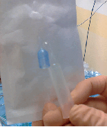
Last summer, Dr. Johansson's office noticed a problem with the phacoemulsification equipment when preparing for a coaxial micro-incision surgery. The operation staff observed that the protection chamber and one silicone sleeve were firmly attached to each other inside the package. The very thin silicone sleeve could have been easily damaged during handling and separation of the two items. In this case, the equipment was exchanged completely, and the observation was reported to the manufacturer. The surgery was uneventful
Source: Bjorn Johansson, M.D.
Although clinical staff must examine surgical instrumentation before use, a recent report recommends taking time to explore one element that tends to be overlooked: the phacoemulsification sleeve.
“In addition to examining the irrigation and aspiration of the phacoemulsification probe, we recommend sparing a few seconds to inspect the phacoemulsification sleeve for leaks or cracks,” according to the study, published online in November 2010 in BMC Ophthalmology.
The phacoemulsification sleeve “tends to be overlooked due to its complementary nature,” the report noted. In the report, a sleeve fracture led to the distal portion remaining inside the anterior chamber upon removal of the phacoemulsification probe.
What happened?
During the case, surgery was performed in the left eye of a 58-year-old woman with grade II nuclear sclerosis and grade I cortical cataract.
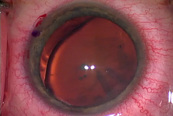
“Towards the end of segment removal, the anterior chamber (AC) was noted to be unstable,” reported lead study author Jennifer W.H. Shum, M.B.B.S., ophthalmology department, United Christian Hospital, Hong Kong. “The followability and aspiration of nuclear fragments also appeared to be impaired. There was a sudden backflow of balanced salt solution emerging from the corneal incision. The AC was still formed at this stage. The surgeon then attempted to withdraw the phacoemulsification probe out of the incision.”
There was resistance to withdrawal, however, as the sleeve was jammed against the cornea.
“This resistance suddenly gave way upon increased traction with resultant severance of the silicone probe sleeve, the distal part of which remained inside the anterior chamber,” Dr. Shum reported.
A surgeon recovered the fragment by first using an OVD to reform the anterior chamber. The retained sleeve was then retrieved with forceps through the corneal incision. The incision did not need to be widened.
No additional smaller sleeve fragments were found in the anterior chamber. The incision was closed with a suture and the patient had uneventful recovery, improving to 20/25 in her left eye at 3 months post-op.
Likely causes
Dr. Shum suggested that a small partial-circumferential fracture of the phacoemulsification sleeve was probably present before surgery. Surgical stress likely extended the fracture.
“This explains the smooth insertion of the probe on commencement of surgery, and the uneventful chopping,” Dr. Shum reported. “At some point, the fracture size was significant enough as to impair the structural turgidity of the sleeve. We believe that the fractured distal part of the sleeve was jammed in the incision. Since the sleeve was elastic, it was possible that the distal part of the sleeve was still in the AC and the break was exposed outside of the incision. This could explain the initial backflow of [balanced salt solution].”
As the phacoemulsification probe was withdrawn, complete severance of the sleeve occurred.
Dr. Shum was unaware as to how the fracture occurred, but suspected it was either something related to the manufacturing process or induced trauma when the phacoemulsification sleeve was applied to the probe by the scrub nurse.
“We therefore advise careful inspection of all instruments before introducing them into the eye,” Dr. Shum concluded.
In the event that sleeve fracture happens in another surgical setting, Dr. Shum advised against using brute force to withdraw the probe.
“If there is resistance on withdrawing the phacoemulsification probe, one can try rotating the probe first, in order to reverse the jam, before attempting to withdraw it again,” Dr. Shum noted. “Excessive force should be avoided to prevent complete severance of the sleeve.”
Bjorn Johansson, M.D., Linkoping University Hospital, Linkoping, Sweden, said that although he has never experienced a fractured phacoemulsification sleeve during surgery, he understands how it could have happened.
“With a decrease in incision size, instruments are getting thinner and more delicate, and that goes for phaco sleeves as well,” Dr. Johansson said. “That means there's a bigger risk for phaco sleeves to tear.”
Dr. Johansson said he has witnessed a phacoemulsification sleeve tear outside of the eye with some amount of manipulation. If a fracture does happen inside the eye, Dr. Johansson, like the authors, suggested it is not insurmountable to get fractured pieces out.
“If you lose a part, it should not be a big challenge to get it out,” Dr. Johansson said. “If it is silicone, I'm not sure that it would be so harmful to the eye.”
Editors' note: Dr. Johansson has no financial interests related to his comments. Dr. Shum has no financial interests related to this study.
Contact information
Johansson: bjorn.johansson@lio.se
Shum: jennishum@gmail.com


留言
張貼留言