What to do? A compromised capsule when a toric IOL was planned
What to do? A compromised capsule when
a toric IOL was planned
EYEWORLD | SEPTEMBER 2023
Precise placement and subsequent stability of a toric IOL are of the utmost
importance for success in correcting astigmatism. Capsule tears can threaten
both factors. So what do you do when a capsule tear occurs during cataract surgery
when you planned to implant a toric IOL? Amandeep Rai, MD, FRCSC, said recognition
is the first step with any case of capsule rent, whether or not a toric IOL is planned.
“Once recognized, the surgeon should try to immediately tamponade the vitreous behind the
compromised capsule with a dispersive viscoelastic device. It is incumbent on the surgeon
to ensure that the anterior chamber remains formed; sudden shallowing may cause the rent
to suddenly enlarge,” Dr. Rai said. “Depending on the stage of the surgery, the surgeon should
attempt to keep all lens material anterior to the rent and remove the cataract with altered fluidics.
Generous use of viscoelastic can help compartmentalize the lens fragments in the anterior
chamber and keep the vitreous posterior. “A surgeon should alter the fluidics by
reducing the flow rate, irrigation pressure, and vacuum,” Dr. Rai continued. “Irrigation and
aspiration may be done manually or at low flow settings. Surgeons should ensure that there is no
vitreous prolapse, and this may be aided by the use of diluted triamcinolone intracamerally. Any
vitreous should be removed using a vitrector, and the surgeon should be vigilant and check
for vitreous regularly through the remainder of the case. Suturing the main wound is suggested,
as this patient may require a vitrectomy and is also at increased risk of postoperative endophthalmitis.
Intracameral antibiotics should also be considered.”
When it comes to IOL selection, Dr. Rai said it depends on capsular support and the type of
rent. If it is an anterior capsule (AC) rent, Dr. Rai said that a single-piece IOL can be placed if
the surgeon is confident in the long-term axial and rotational stability.
“This depends on appropriate placement of the haptics so that a haptic does not tilt
forward; if a single haptic is in the bag and the other haptic tilts forward into the sulcus, the
patient is at high risk for postoperative uveitisglaucoma- hyphema (UGH) syndrome,” he said.
“The tilt can also induce astigmatism and/or coma. The ideal scenario would be a small AC
rent that happens to coincide with the steep axis of corneal astigmatism. This would allow
the physician to orient the toric IOL such that the haptic-optic junction is around the area of
the rent, and both haptics will be entirely secured under the remaining capsulorhexis edge.”
A single-piece IOL may also be an option in the setting of a posterior capsule rupture (PCR),
provided the posterior capsule surface area is large enough to support the IOL long term.
“If the surgeon is able to keep the PCR small and controlled throughout the remainder
of the surgery, a single-piece IOL is certainly a plausible outcome; in that scenario, a toric IOL
should be considered,” he said. “This is especially true if the PCR can be converted to a posterior
continuous curvilinear capsulorhexis. If the PCR is large and the surgeon thinks that the
remaining posterior capsule cannot support an IOL, a common IOL placement is in the sulcus
(with or without optic capture). In this scenario, the surgeon should not place a single-piece toric
IOL in the sulcus due to the increased risk of UGH syndrome. Instead, a three-piece IOL with
PMMA haptics should be used. An alternate option for placement of a single-piece IOL in
the setting of a PCR is reverse optic capture; a single-piece IOL can be placed with the haptics
in the capsular bag and the optic anterior to the capsulorhexis opening. This technique of
reverse optic capture would allow a surgeon to still implant a single-piece toric IOL in a compromised
capsule.”
If capsular support is entirely insufficient Dr. Rai also mentioned anterior chamber IOLs,
iris-sutured IOLs, scleral-sutured IOLs, and intrascleral haptic fixation as possibilities.
Jonathan Rubenstein, MD, shared his thoughts on what to do when there is a compromised
capsule and a toric IOL was planned. If there is a PC tear, he said you need to make
sure you can visualize the entire extent of the tear to ensure that it won’t tear out, producing
instability. If it’s localized (and ideally round), Dr. Rubenstein said it’s unlikely to tear out,
and thus OK to place a toric IOL, provided the zonules are still good.
“During placement, avoid further extension of the posterior capsule, using OVD to protect
the capsular bag,” he said. Dr. Rubenstein said if it’s not advisable to
place a toric IOL, you can still address astigmatism in the OR. If you’ve planned for it or have a
nomogram and the proper equipment available, you could perform limbal relaxing incisions
(LRIs), he said. He added that he doesn’t think many surgeons are comfortable or have the
equipment/information available to them in the OR to perform this procedure, if they weren’t
already planning for it. Postop management of astigmatism, if a
monofocal IOL was placed due to the compromised capsule, includes glasses, toric contact
lenses, or a refractive procedure, such as corneal refractive surgery, LRIs, astigmatic keratectomy,
and opposite clear corneal incision. Dr. Rubenstein said these are options for patients who had
a three-piece lens in the sulcus or placed with optic capture. He lets these patients stabilize
for 3 months post-cataract surgery because “at that point, it’s refractive astigmatism rather than
astigmatism based on corneal measurement.” The physicians also addressed the patient
counseling aspect of this complication. “The discussion,” Dr. Rubenstein said, “is:
‘Our first priority is to get your cataract out safely and completely, which we were able
to accomplish. … Second, we want to put a lens implant in your eye that is as close to the
correct power and as stable as possible, and we were able to accomplish that. Third is to try to
produce the lowest residual refractive error … as possible, and we’re able to correct hopefully
the spherical part of your refraction, but you still have astigmatism, which we were not able
to correct in surgery, and we will offer you the opportunity to correct that later.’ We’ll say something
like, ‘During surgery we assessed that your eye was not stable enough to support the type
of lens implant that we originally had planned to correct astigmatism; we thought it was unsafe
to use that kind of lens because we couldn’t be assured it would stay in the position that was
needed to fully correct your astigmatism, and therefore we put in a lens implant that does not
correct astigmatism because it was the most stable lens for your eye. We can always come back
later and correct your astigmatism.’” Dr. Rai also said it’s important to thoroughly
discuss this situation with the patient and theirfamily postop, namely because these patients
are at increased risk for complications, such as high intraocular pressure in the first few hours
postop, endophthalmitis in the days postop, and/or retinal tear/detachment, CME, or
pseudophakic bullous keratopathy in the weeks postop.
“They may also need further surgery for retained fragments. As such, patient education
is important so they may seek immediate and appropriate care for any postoperative complications.
These patients should also be scheduled for close follow-up to monitor for complications
and ensure a safe recovery,” Dr. Rai said. Overall, Dr. Rai said that patients with
astigmatism can benefit from a well-placed toric IOL, and even in the setting of some capsular
complications, it is still possible, depending on a few factors, to deliver the best possible uncorrected
distance visual acuity to patients.

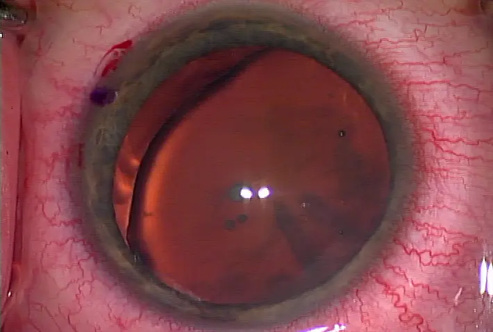
A toric IOL on axis, placed after a localized posterior capsular tear
Source (all): Jonathan Rubenstein, MD


留言
張貼留言