Vitrectomy prior to cataract removal for the "crowded anterior segment"
Complicated cataract cases
Vitrectomy prior to cataract removal for the "crowded anterior segment"
by Samuel Masket, M.D., Basak Bostanci Ceran, M.D., and Nicole R. Fram, M.D.
 Eyes with very shallow anterior chambers come along every now and then. Most are anatomically small in the anteroposterior dimension and carry hyperopic refractive errors. The central anterior chambers of these eyes may be only a few corneal thicknesses deep, and their irises may be in apposition to the peripheral posterior cornea. Often they have had a peripheral iridectomy already because they are anatomically predisposed to phakic pupillary block and angle closure glaucoma. Despite these obstacles, most of these eyes can undergo fairly routine cataract surgery with a few extra considerations—a little pre-op digital massage, the administration of a hyperosmotic agent, a carefully constructed cataract incision that prevents iris prolapse, and the judicious use of a highly retentive OVD. Every now and then, however, these relatively simple measures are not enough. Some times the patient has severe pain and an intraocular pressure of 60 mm Hg on presentation. The eye is in acute angle closure, or worse yet, phacomorphic or rubeotic angle closure. The anterior chamber may only be the depth of the pupil sphincter. In such eyes, there may be no anterior chamber into which to make a paracentesis. The iris may be against the posterior cornea. Once made, the iris prolapses immediately into the paracentesis incision. Forced entry with an OVD cannula only causes iris trauma. These are the eyes in which it might be necessary to perform a dry vitreous tap and make room in the posterior segment for the lens to drop back. It is a trick any cataract surgeon can learn. In their article, Drs. Masket, Ceran, and Fram describe their experience with the dry vitreous tap technique for the rare eye that presents with a very crowded anterior segment.
Kevin Miller, M.D., complicated cataract cases editor


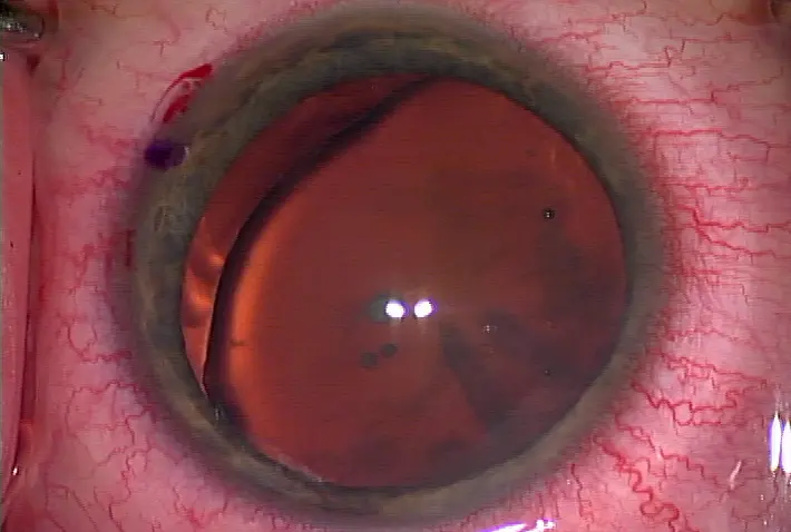
Figure 2 Source: Samuel Masket, M.D.
On very rare occasions, anterior chamber depth is inadequate to allow for cataract surgery to be safely performed. Ancillary measures, including digital ocular massage and intravenous administration of mannitol, in combination with the use of a viscoadaptive OVD, are generally sufficient to manage the "crowded anterior segment." However, should anterior chamber depth be inadequate to perform capsulorhexis and other stages of surgery, the cataract surgeon may need to consider a very limited pars plana vitrectomy prior to cataract surgery. In this infrequent scenario, the surgeon would note that the globe becomes "rock hard" after anterior chamber instillation of a viscoadaptive OVD, yet the chamber depth remains inadequate for cataract surgery. Intumescent mature cataract and anterior segment microphthalmos are typical causes. Although the anterior chamber may be shallow in cases of diffuse zonulopathy, the chamber will often deepen on administration of an OVD.
The surgical technique involves making a single pars plana entry with a disposable MVR blade, generally 3.5 mm posterior to the limbus. The unsleeved vitrectomy probe is aimed toward the middle of the globe and angled posteriorly in order to avoid damage to the posterior lens capsule; no infusion is necessary. High-speed cut rate and low vacuum setting is carried out for only a few seconds, the probe carefully removed, and the anterior chamber deepened with additional OVD. In this manner, the vitreous volume is reduced, the anterior chamber deepened, and ocular hypotony is avoided. Varying with the nature of the sclerotomy, suture closure may be necessary at this time.
The practice of pars plana vitreous aspiration at cataract surgery in this clinical scenario has been described previously.1 In fact, during the infancy of lens implant surgery, and before the advent of OVDs, not infrequently surgeons would employ pars plana needle aspiration of vitreous in order to create anterior chamber space for lens implantation. With the advent of automated vitrectomy, the technique changed and was eventually published in the peer-reviewed literature by David F. Chang, M.D.2 Two following cases are illustrative.
Case 1
A 69-year-old male was referred following an attack of acute angle-closure glaucoma. An intumescent, mature cataract ensued and cataract surgery was attempted. However, at surgery the chamber was noted to be extraordinarily shallow and the procedure aborted prior to anterior capsulotomy.
Upon examination one could note a prior superior iridectomy, posterior synechiae with an irregular, poorly dilating pupil, and an associated pupillary membrane (Figure 1). The cataract was fully white, precluding examination of the posterior pole. However, the patient had brisk two-point light discrimination and grossly normal color perception. B-scan ultrasonography revealed a normal posterior segment. The fellow eye was pseudophakic, but otherwise normal to examination. At surgery the patient received peribulbar injection anesthesia, digital ocular massage, and 50 grams of intravenous mannitol. Despite these maneuvers, instillation of a small amount of DisCoVisc (Alcon, Fort Worth, Texas) via a paracentesis was accompanied by marked elevation of IOP, yet chamber workspace was inadequate. As described, an automated pars plana vitreous aspiration was performed and surgery completed without complication. Surgery was complex in that it was necessary to perform synechiolyis, pupil membranectomy, pupil stretch, and trypan blue capsule staining. As the cataract was intumescent, copious liquefied white cortical material effused upon initiation of the capsulorhexis, but the capsulotomy was safely completed in two stages after aspirating the material (Figure 2). It is quite likely that by allowing the anterior capsule to flatten with OVD, the vitrectomy prevented uncontrolled tearing of the anterior capsule, in the manner of the "Argentinian flag sign".
The post-op course was routine and the patient returned to normal visual function. However, owing to pre-existing synechiae, the pupil remained slightly irregular post-op.
Case 2
A 68-year-old female came under care in 2005 and was noted to have asymmetric anterior chamber depths, with the LE noted to be particularly shallow. This eye had a previous laser peripheral iridotomy. Moreover, she had pseudoexfoliation, although there was no evidence of phacodonesis. Gradually, the involved eye developed a visually significant cataract. Presurgical testing revealed an anterior chamber depth of less than 2.0 mm.
At surgery the chamber could not be adequately deepened with addition of DisCoVisc. Following administration of a small amount of subconjunctival anesthetic, a conjunctival peritomy was created and a limited automated pars plana vitrectomy performed as described above. Once completed, surgery was carried out routinely. Interestingly, there was no evidence of zonular weakness, although a capsular tension ring was placed prophylactically. Fortunately, in this case, the vitrectomy probe could be visualized, reducing the risk of a "blind vitrectomy," as was needed for case 1. Her post-op course was routine.
Although very rarely indicated, limited, single-port pars plana automated vitrectomy may be necessary to safely perform cataract surgery in a crowded anterior segment. Concomitant with or just afterward, the anterior chamber may be deepened with OVD and hypotony averted. Only small amounts of vitreous need to be removed in this technique. The overall concept is to maintain normal IOP while expanding the anterior chamber by reducing posterior segment volume.
References
1. Machemer R. Symposium: pars plana vitrectomy. Introduction. Trans Am Acad Ophthalmol Otolaryngol 1976; 81:350-351.
2. Chang, DF. Pars plana vitreous tap for phacoemulsification in the crowded eye. J Cataract Refract Surg 2001; 27:1911-1914.
Editors' note: Drs. Ceran, Fram, and Masket have no financial interests related to their comments.
Contact information
Ceran: basakbostanci@hotmail.com
Fram: nicfram@yahoo.com
Masket: sammasket@aol.com


留言
張貼留言