The role of corneal topography in premium-channel cataract surgery
The Pentacam:
Offering a Clearer View
The Pentacam Comprehensive Eye Scanner (Oculus, Inc., Lynnwood, WA) continues to expand the field of ophthalmic diagnostics with new features and software, such as densitometry for cataracts. This and other key information on the Pentacam is presented in the following articles, which summarize presentations that were given at two breakfast symposia on November 10th and 11th in New Orleans.
SUPPLEMENT TO CATARACT & REFRACTIVE SURGERY TODAY I FEBRUARY 2008
Surgical Refinement
The role of corneal topography in premium-channel cataract surgery.
BY JOHN A. VUKICH, MD
I n this visual age, nervous patients are often reassured when they can see an image or set of data that shows (1) you are a capable surgeon and (2) that you can identify and treat their visual problems. The Pentacam Comprehensive Eye Scanner (Oculus, Inc., Lynnwood, WA) is an educational aid and a diagnostic workhorse that fulfills this function in my practice. Here, I describe how my staff and I use this device on a daily basis.
ASTIGMATIC CORRECTION
Refractive cataract surgery is here to stay. To succeed at it, however, we need to continuously refine our techniques and meet our patients’ expectations for spectacle-free postoperative outcomes. For most of us, transitioning to this new treatment paradigm requires adopting an approach to patients’ education and preoperative work-up that is different from what we learned as residents. Because many refractive cataract patients undergo an additional procedure to achieve spectacle freedom, we must be willing to correct residual spherical and astigmatic errors. Astigmatism is common; approximately 40% of patients have 1 or more diopters of preexisting astigmatism.1 How common is keratoconus? The general consensus is that the condition occurs in one in 2,000 patients, although the rate is higher in those who have preexisting astigmatism. Thus, the ability to detect and diagnose keratoconus preoperatively is becoming increasingly critical so that we may discuss appropriate treatment options with the patient. Such detection is particularly important when we are considering implanting a premium-channel IOL, because none of these lenses currently corrects astigmatism.
CASE STUDIES
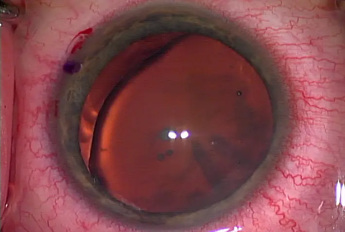
Case 1 Because multifocal lenses introduce significant aberrations into the visual system, the second-order aberrations (sphere and cylinder) must be corrected, or else they will compound the higher-order aberrations inherent in multifocal technology. As little as 1.50 D of residual error can be bothersome to patients. A 52-year-old patient with normal preoperative topography underwent implantation in his left eye with theReZoom IOL (Advanced Medical Optics, Inc., Santa Ana, CA) that produced a UCVA of 20/50, +0.75 D spherical equivalent, and +1.50 D of sphere. The patient’s wavefront point-spread function showed significant visual irregularity that correlated with his complaints of starbursts and halos. Although I had wanted to treat his residual error with a customized wavefront ablation, his wavefront reading did not correlate with his manifest refraction (Figure 1). Thus, he underwent a standard IntraLASIK procedure (IntraLase Corp., Irvine, CA) to correct his residual errors and achieved a manifest refraction of plano and a UCVA of 20/20.

Case 2 Topography is crucial to refractive cataract surgery, and surgeons who wish to adopt premium-channel IOLs must become comfortable with using it routinely. Increasingly, the cataract population is encompassing younger patients who expect a refractive outcome as part of their surgery. Suddenly, cataract surgeons must assess such items as corneal contour and refractive stability in order to choose the best IOL for their patients. Figure 2 shows a 48-year-old cataract patient with brightness acuity testing readings of 20/100, a BCVA of20/30, and 2.00 D of cylinder. The Pentacam showed an abnormal inferior/superior ratio, indicating an atypical anterior segment. This patient was contraindicated for a corneal relaxing incision and a multifocal IOL. Fortunately, the Pentacam provides insights for treating patients with limited options. Its algorithms help us tease out a patient’s risk for surgical complications. I decided to treat him with a toric IOL. Postoperatively, his manifest refraction was plano, and his UCVA was 20/25. Considering that his cornea was irregular preoperatively, this patient feels that his vision is much improved, and he is extremely happy. Although he requested a multifocal IOL when he presented, the Pentacam allowed me to select a more appropriate treatment for this patient.

IOL CALCUL ATION AND SELECTION WITH THE PENTACAM
We now have access to a variety of IOLs, including toric and aspheric aberration-adjusted lenses. In order to choose the best lenses for patients and provide optimal outcomes, we need to understand what causes corneal aberrations and how to prevent and treat them, if necessary. Positive and negative spherical aberration are derived from corneal topography. Therefore, cataract surgeons who plan to implant IOLs need to know the location of the corneal aberration they are trying to correct and which of the four available IOLs will produce the best result. I believe that intelligently selecting an IOL based on preexisting phenomena and other considerations amounts to customized cataract surgery. The Pentacam provides all of this information in its Holladay Report and elevation and pachymetry maps. In fact, the system’s next software upgrade will match a patient’s root mean square on the Zernike display and give the specific information. Choosing an IOL for an eye that previously underwent LASIK, PRK, or RK requires special consideration. We cannot rely on standard keratometry to calculate the proper IOL power, because our corneal model assumes that the positive anterior surface and negative posterior surface occurs in a fixed ratio. Because LASIK alters this relationship, we cannot plug keratometric measurements taken from postLASIK corneas into standard IOL formulas without performing additional recalculations. Increasingly, we require a tool to accurately predict a patient's net corneal power (effective corneal power), which will allow us to predict an implant's power. I use the Holladay Report available on the Pentacam to more accurately calculate IOL power for my postrefractive patients. I think the algorithm is outstanding, and it has changed the way we approach these IOL calculations. The report calculates the cornea's front-to-back ratio power using true power calculations before generating the neteffective power based on the anterior and posterior surfaces. These K readings reflect the true optical power of the cornea following refractive surgery and can be used with any of the standard IOL power calculation formulas. The Holladay Report also calculates relative pachymetry. Jack Holladay, MD, MSEE, studied a database of over 1,000 normal corneas and determined their average thickness. The Pentacam calculates the degree to which the cornea being measured deviates from this average over the entire surface of the cornea. Deviations of 10 µm or greater in any area tend to indicate an abnormality and help identify suspicious patterns that can indicate a potential problem.
CONCLUSIONS
All corneal and cataract surgery must be performed on stable corneas, and I believe that the Pentacam is an important tool to help surgeons identify corneal abnormalities and thus improve our services. The system’s ease of use and reliability have increased my efficiency. I no longer spend hours looking at topography; the Pentacam’s printouts allow me to make a surgical decision quickly and accurately. It has improved my outcomes by helping me identify the best surgical treatments for my patients.
■ John A. Vukich, MD, is Surgical Director of the Davis Duehr Dean Center for Refractive Surgery in Madison, Wisconsin. He is not a paid consultant for Oculus, Inc., and acknowledges no financial interest in the company or its products.
Dr. Vukich may be reached at (608) 282-2000; javukich@hotmail.com. 1. Grabow HB, Martin RG. STAAR AA-4203 one-piece plate-haptic silicone IOL. In: Martin RG, Gills JP, Sanders DR, eds. Foldable Intraocular Lens. Thorofare, NJ: Slack, Inc.; 1993: 237-250.


留言
張貼留言