Challenging refractive cases A case of isolated epithelial ingrowth nests?
August 2010‧EyeWorld
Challenging refractive cases A case of isolated epithelial ingrowth nests?
by Michelle Dalton EyeWorld Contributing Editor
Frustration mounts as a recurrent epithelial ingrowth is evasive to treat
It all seems to go according to plan—the patient is evaluated for LASIK, is determined to be a good candidate, the surgery scheduled, the microkeratome-created flap and subsequent laser correction are completely uneventful, immediate post-op indicates this is yet another successful surgery. But then the woman returns a couple of months later with an epithelial ingrowth. Instinct tells the surgeon to lift and scrape and that will be that. About 6 weeks later, however, the woman returns with an epithelial ingrowth in just about the same spot as the original. Again, the surgeon presumes he might have missed a cell or two the first time, so he lifts, scrapes, replaces one more time. He notes the epithelial cells seem to be very central, with no visible trail. Yet what should the surgeon do if she presents one more time? That was the dilemma facing Jay "J.E." McDonald, M.D., Fayetteville, Ark., editor of the ASCRS Cataract and Refractive Internet Forum, recently. Dr. McDonald agreed to allow EyeWorld to present this complicated case, in hopes that other surgeons might learn from his experience.
 |
| by Michelle Dalton EyeWorld Contributing Editor |
 |
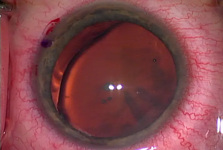
Newly formed epithelium after PRK is not always firmly attached to the cornea Source: Dajlit Singh, M.D. |
Dr. McDonald said one of the more frustrating aspects of managing this case was that the ingrowth appeared to return in almost the exact spot he found it initially. "The first time she presented with the ingrowth, I lifted and scraped the top and bottom of the flap aggressively to remove what I consider all the 'slimy' cells," he said. "Because they were so close to the visual axis, I was a little reluctant to get overly aggressive in case I created an inadvertent tear or cut the fibers." He added a downside to lifting and scraping is that "you get swelling and haze. That can make it very difficult to see the cells."
Culture the cells
In general, what makes this case difficult and unexpected is that the incidence of epithelial ingrowth after primary LASIK is low, "especially with the newer microkeratomes," said Sonia H. Yoo, M.D., associate professor of clinical ophthalmology, Bascom Palmer Eye Institute, Miller School of Medicine, University of Miami, Miami. "The edges of the side cut are more precise, and there's less of an acute angle." Likewise, the femtosecond laser creates a much steeper angle during flap creation; in both cases, "visually significant primary epithelial ingrowth is less common these days," Dr. Yoo said. "I've done LASIK for 12 years or 15 years now," Dr. McDonald said, "and I've used every microkeratome around, and so I had some epithelial ingrowth back in the day. This particular case was just very unusual in terms of where the cells presented and how they presented."
Dr. Yoo agreed, noting there are typically two types of epithelial ingrowth variants—isolated clusters of cells underneath the flap "that are implanted inadvertently" and those that appear "like a peninsula from the edge of the flap." The latter occurs when epithelial cells get underneath the flap edge and continue to grow from there. Isolated nests as Dr. McDonald encountered "don't tend to progress," Dr. Yoo said, noting the cells need a connection to the outside in order to grow.
Epithelial ingrowth can be visually significant if (or when) it reaches the center, Dr. Yoo said. Cells that are connected to the peninsula "tend to progress over time; they'll progress slowly, but they will progress," she said. "If you have an ingrowth that's growing rapidly, either its connection is broad from the outside or the stalk is wide and lets more cells in that encourages its growth."
In the latter case, though, surgeons also need to consider the timing of the ingrowth presentatio—if the ingrowth begins to develop within the first week post-op, "one should be suspicious for infection," Dr. Yoo said. "If you're scraping, you might as well culture because you might be fooled about what it is," she said, and as long as the flap is being lifted, culturing the cells makes sense. For those practices without access to plates, tubes can work well for this type of situation. Another way to differentiate between an ingrowth and an infection is to use confocal microscopy (if available); epithelial ingrowth cells have an unusual appearance that's not likely to be confused with anything else. Dr. Yoo said epi ingrowth is "very typical with a stalk connected to the edge of the flap, and a whirl-like pattern with a clear leading edge just in front of the opaque portion of the ingrowth." The acknowledged problem with epi ingrowth is how difficult eradication may be. She suggested removing a "little bit" of the epithelium on the outside of the flap, to ensure breaking the connection between the outside and underside of the flap. Then she'd use bandage contact lens, antibiotics and steroids until the epithelium had healed. "If there's any question of infection, I may use a broader coverage antibiotic," she said. If the initial lift and scrape did not work, surgeons can re-lift, but may have to place sutures, Dr. Yoo said. "The disadvantage to that is you might induce astigmatism, but the technique is fairly effective at reducing the chance of recurrence."
She said surgeons can also try sealing the flap with bioadhesive glue, but said a disadvantage is they may be bulky "and you'd have to cover the flap with the bandage contact lens, so the contour isn't really smooth." If given a preference, Dr. Yoo prefers suturing before sealing with glue. Using alcohol—"that's toxic to the epithelial cells but not to other cells"—could be considered. Mitomycin-C "can be used, and might be effective. You could try to do adjunctive PTKon the bed to eliminate any residual cells as well," Dr. Yoo said.
Try something new
Patient age can be a risk factor for epithelial ingrowth, said Elizabeth A. Davis, M.D., adjunct assistant professor of ophthalmology at the University of Minnesota, Twin Cities, Minneapolis. Up to 20% of older patients may have anterior basement membrane dystrophy (ABMD), and they may be better candidates for PRK than LASIK, even if a femtosecond laser flap is part of a surgeon's technique.
"With microkeratomes, there's a translational force across the flap, and that can abrade loose cells off the flap," she said. "That can lead to epithelial ingrowth, localized diffuse lamellar keratitis, poor healing, poor vision, etc."
Subtle ABMD can sometimes be difficult to correctly diagnose, she added, and suggests surgeons "widely dilate the pupil and look for the characteristic maps, dots, and fingerprints." Corneal topography can also help alert surgeons to the irregular astigmatism that frequently occurs in this condition, she said. A focal—and very tiny—buttonhole could be the source of the recurrent cells in Dr. McDonald's case, Dr. Davis said. "If there's even the smallest buttonhole, lifting and scraping may not be successful, as there's still a way for the cells to migrate in," she said.
If the first lift and scrape does not eradicate the ingrowth completely, Dr. Davis suggests trying something different when the first recurrence presents. In this approach, prior to lifting the flap, she recommends de-epithelializing the whole cornea "to within a millimeter of the limbus." "I'd use diluted alcohol until all the cells loosen and then debride," Dr. Davis said. "Go over the flap and beyond the flap edge, 360-degrees. Once that's all cleared off, I'd try to identify any difference in the corneal reflex in the area of the ingrowth to identify a possible buttonhole through Bowman's membrane. Following that, I'd lift the flap and remove the epithelial cells from the stromal bed and back of the flap carefully. Then I'd take a little bit of 70% alcohol on a sponge and dab it where the epi nest was to lyse any stray cells as these can be the source of another recurrence." Lastly, Dr. Davis would replace the flap, irrigate beneath it, stretch it back into position and apply fibrin glue (Tisseel, Baxter Corp., Mississauga, Ontario) to the entire cornea. The glue takes about 8 minutes to polymerize and "then I place a bandage contact lens. The near total de-epithelialization and fibrin glue allow an extended period of time for the flap to adhere" without the presence of any epithelial cells in the area. "You should leave the bandage contact lens on for about 2 weeks while the Tisseel glue dissolves," she said.
If the ingrowth is truly recurring as isolated nests with no connection to the flap edge, suturing may not be successful, Dr. Davis said. "If there's a tunnel or a track extending to the flap edge, you could suture it and seal down the track line. YAGing the epithelial nest is an option as well," she added.


留言
張貼留言