Obtaining successful surgical outcomes for patients with posterior polar cataracts
August 2011‧Eye World
Obtaining successful surgical outcomes for patients with posterior polar cataracts
by Uday Devgan, M.D., F.A.C.S., F.R.C.S.
Dr. Devgan, Devgan Eye Surgery, Los Angeles, and Beverly Hills, Calif., details his approach to cataract surgery for patients with posterior polar cataracts
 |
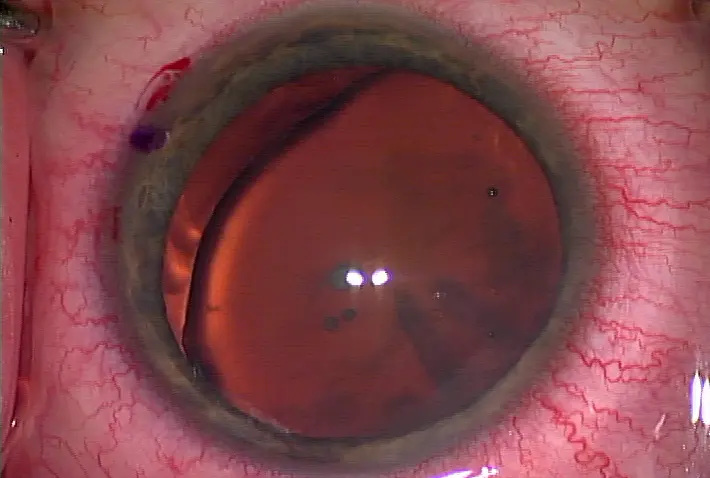
This patient has a posterior polar cataract with a congenital iris defect and absence of zonules in one quadrant Source: Uday Devgan, M.D., F.A.C.S., F.R.C.S. |
Phacoemulsification of a posterior polar cataract is particularly challenging because the posterior capsule can be weak, fragile, or even absent at the site of the opacity. This leads to a higher risk of posterior capsule rupture, vitreous loss, and other complications. Initial studies have demonstrated that this risk is as high as 26%2 to 36%3 although recent reports peg the risk a bit lower at 11%.4 The size of the posterior polar opacity may also influence the risk of capsular rupture, with larger opacities thought to have a higher risk.5
Multiple surgical techniques and approaches have been described, but the one common goal is to avoid manipulation of the posterior capsule at the site of the posterior polar opacity. The technique that I prefer is hydrodelineation to remove the central lens endonucleus, followed by viscodissection of the remaining lens epinucleus and cortex.6,7
The surgical technique
Creation of a well-centered, 5-mm, round capsulorhexis is particularly important in an eye with a posterior polar cataract since there is a significant chance that the IOL will need to be placed in the ciliary sulcus with optic capture through the anterior capsular opening.
It is important to avoid performing hydrodissection near the posterior lens opacity, since the fluid wave can cause the posterior capsule to rupture and the nucleus to fall into the vitreous. While some surgeons advocate a small amount of hydrodissection, stopping just shy of the posterior pole, my advice is to avoid this step altogether.
Hydrodelineation using a small quantity of balanced salt solution in a syringe with a 27-gauge cannula can be performed since this will separate the endonucleus from the remaining epinucleus and cortex. The endonucleus can then be removed from the eye using the phaco probe.
At this point, all that remains in the capsular bag is the softer lens epinucleus and the cortex. Using a dispersive viscoelastic, which has a more liquid and syrup-like texture than the cohesive viscoelastics, the remaining lens material can be carefully dissected from the capsule. Using a viscodissection technique in all quadrants of the lens allows for a complete cleaving of all residual lens material from the capsule. This method has several benefits: the viscoelastic is slow and controlled, it pressurizes the anterior segment, it can tamponade any existing break in the capsule, and it creates a barrier between the lens material, which is brought forward, and the capsule and vitreous, which are pushed backward.
The irrigation/aspiration probe can be placed in the eye and kept centrally in the anterior segment while the lens material is aspirated. The risk of a capsular rupture is highest during attempted manipulation or cleaning of the posterior polar opacity. While the posterior polar opacity can often be removed from the capsular surface, care should be taken to avoid polishing or cleaning. It is far easier and less risky to perform a YAG laser capsulotomy to clear the visual axis in the post-op period.
Inserting the IOL
When the lens material has been removed, it is critical not to let the anterior chamber collapse. This means keeping the I/A probe in the eye in foot pedal position 1 to maintain the infusion pressure and then using the non-dominant hand to inject viscoelastic via the paracentesis incision to fully inflate the capsular bag. At this point the I/A probe can be removed from the eye and the new IOL inserted.
A three-piece IOL is preferred since it offers more options for placement. The entire IOL can be placed in the capsular bag if the posterior capsule is intact. If a defect of the posterior capsule develops during IOL insertion, the haptics can be placed in the sulcus with the optic captured via the buttonhole technique through the capsulorhexis. This provides excellent stability of the lens and creates a barrier to prevent vitreous prolapse.
The post-op course for these patients tends to be straightforward, particularly if the posterior capsule remains intact. After contraction of the capsular bag has created a strong fixation for the IOL, a YAG laser capsulotomy can be performed for any residual posterior capsule opacity. Using viscodissection, posterior polar cataracts can be effectively treated while minimizing the risks.
References
1. Addison PK, Berry V, Ionides AC, Francis PJ, Bhattacharya SS, Moore AT. Posterior polar cataract is the predominant consequence of a recurrent mutation in the PITX3 gene. Br J Ophthalmol. 2005 Feb;89(2):138-41.
2. Osher RH, Yu BC, Koch DD. Posterior polar cataracts: a predisposition to intraoperative posterior capsular rupture. J Cataract Refract Surg. 1990;16:157–62.
3. Vasavada A, Singh R. Phacoemulsification in eyes with posterior polar cataract. J Cataract Refract Surg. 1999;25:238–45.
4. Lee MW, Lee YC. Phacoemulsification of posterior polar cataracts—a surgical challenge. Br J Ophthalmol. 2003 Nov; 87(11): 1426–1427.
5. Kumar S, Ram J, Sukhija J, Severia S. Phacoemulsification in posterior polar cataract: does size of lens opacity affect surgical outcome? Clin Experiment Ophthalmol. 2010 Dec;38(9):857-61.
6. Fine IH, Packer M, Hoffman RS. Management of posterior polar cataract. J Cataract Refract Surg. 2003 Jan;29(1):16-9.
7. Devgan U. Visco-Dissection Technique for Posterior Polar Cataracts. Grand Prize winner, Asia-Pacific Academy of Ophthalmology Meeting 2005, Kuala Lumpur, Malaysia.
Editors' note: Dr. Devgan has no financial interests related to this article.
Contact information
Devgan: 800-337-1969, www.devganeye.com


Thanks to you for published this great post.
回覆刪除Eye Surgeon in Bristol
Cataract Surgeon in Bristol
Ophthalmologist in Bristol
https://www.momajid.com/eye-care.php?premium-cataract-surgery-in-bristol&id1=6