"Inside-out" approach to posterior polar cataracts
"Inside-out" approach to posterior polar cataracts
by Abhay R. Vasavada, M.S., F.R.C.S., and Shetal M. Raj, M.S.

Although most posterior polar cataracts tend to be on the softer side at the time of their removal, they can still be some of the more complex cataract extractions. The thinned and weakened posterior capsule has been reported to rupture in 26-36% of these cases. Certain precautions can be taken such as avoiding downward pressure on the capsule, avoiding hydrodissection, avoiding excessive rotation of the endonucleus, and working within the safety of an epinuclear shell. In this month's column, one of the world's most experienced surgeons, Abhay Vasavada, M.S., F.R.C.S., gives tips and pearls for dealing with the posterior polar cataract. His inside-out hydrodelineation technique is a useful method for insuring safe creation of an epinuclear shell while placing minimal stress on the posterior capsule. His step-by-step instructions should help us all lower our rate of capsule rupture in these difficult cases.

Figure 1. A moderate-sized continuous curvilinear capsulorhexis is created

Figure 2. A central trench is sculpted using the slow motion technique

Figure 3. A dispersive viscoelastic is injected through the side port before retracting the probe to maintain a closed chamber

Figure 4. A specially designed right-angled cannula is introduced through the main incision to reach the central trench

Figure 5. The tip of the cannula is placed adjacent to the left wall of the trench at an appropriate depth

Figure 6. Fluid is injected slowly with minimal force through the left wall of the trench Source (all): Abhay R. Vasavada, M.S., F.R.C.S., and Shetal M. Raj, M.S.

Figure 7. Delineation is produced by the fluid traversing inside-out

Figure 8. If the ring is incomplete, fluid injection may be repeated in the right wall of the trench with another right-angled cannula facing left

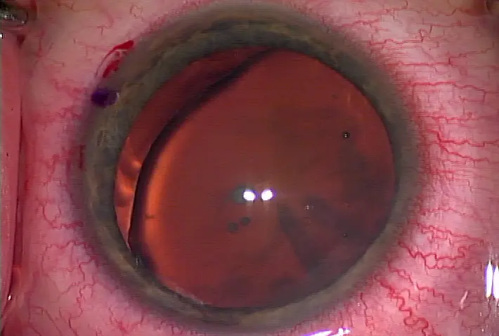
Figure 9. A well-demarcated ring within the lens denotes the endpoint of delineation

Figure 10. A precise demarcation of the central core nucleus from the epinucleus provides a thick epinucleus bowl

Figure 11. A sketch to demonstrate inside-out delineation Source (all): Abhay R. Vasavada, M.S., F.R.C.S., and Shetal M. Raj, M.S.
Posterior polar cataracts pose a unique challenge to the surgeon owing to the high risk of posterior capsule dehiscence during emulsification. It has been observed that the risk of posterior capsule rupture (PCR) increases with performing conventional cortical cleaving hydrodissection. The speculation is that injecting balanced salt solution in the capsular bag with an intact nucleus rapidly builds up the hydraulic pressure within the bag. This pressure is adequate to split the capsule at the site of the polar opacity as fluid passes through the area of least resistance. Other risk factors that could cause a PCR in eyes with posterior polar cataracts (PPC) are sudden and excessive forward movement of the iris-lens diaphragm, premature cleavage of the opacity from the capsule, or attempts to polish the central plaque opacity on the posterior capsule. To circumvent this eventuality and to facilitate nucleus removal during phacoemulsification in eyes with posterior polar cataract, we devised the inside-out delineation approach.
Pre-procedure evaluation
PPC has to be distinguished from the posterior subcapsular cataract. PPC is characterized by a central, dense, disk-shaped opacity located on the posterior capsule with concentric rings around the central plaque opacity that appear like a bull's eye. The opacity has a cone-shaped projection in the subcapsular region or central posterior cortex. The posterior subcapsular cataract is rather flat and does not have distinctly demarcated central ring opacity. PPC could also have co-existing nuclear or cortical opacities. Noting the integrity of the posterior capsule could help explain the prognosis to the patient.
Rationale
It is widely accepted that cortical cleaving hydrodissection should be avoided during emulsification of posterior polar cataracts as it increases the risk of posterior capsule rupture. Conventional hydrodelineation is instead performed, wherein the cannula is penetrated within the lens substance in an attempt to cause the fluid to traverse between the nucleus and epinucleus. With this there remains a possibility of fluid being injected inadvertently between the opacity and the capsule, leading to unwarranted hydrodissection. Also at times it may be difficult to introduce the cannula within a firm nucleus leading to rocking and stress to the capsular bag and zonules. With inside-out delineation fluid is injected at a desired depth, under direct vision. A precise demarcation of the central core nucleus from the epinucleus provides a thick epinucleus bowl. This bowl acts as a mechanical cushion and a barrier that protects the posterior capsule during subsequent maneuvers.
Instrumentation, anesthesia, and technique
A. Instrumentation • Operating microscope with good coaxial illumination • Dispersive viscoelastic: I prefer chondroitin sulfate (Viscoat, Alcon, Fort Worth, Texas).
• A specially designed 27-gauge right-angled cannula facing the right and the left side B. Anesthesia • Peribulbar anesthesia is advisable for novice surgeons; with experience topical anesthesia could be administered. C. Technique • A moderate-sized continuous curvilinear capsulorhexis is created (Figure 1). No hydro procedure is done, and the surgeon commences phacoemulsification. A central trench is sculpted using the slow motion technique (Figure 2). In nuclear sclerosis less than grade 3 (grading system from grades 1-5): • Ultrasound energy (U/S): 30-40% • Vacuum: 40 mm Hg • Aspiration flow rate (AFR): 14 cc/min • Bottle height: 50 cm Care should be taken not to mechanically rock the lens. • Dispersive viscoelastic is injected through the side port before retracting the probe to avoid forward movement of the iris-lens diaphragm (Figure 3).
• A specially designed right-angled cannula mounted on a 5 cc syringe filled with balanced salt solution is introduced through the main incision to reach the central trench. (Figure 4). The tip of the cannula is placed adjacent to the left wall of the trench at an appropriate depth (Figure 5). The plane at which the cannula penetrates the wall of the trench (buried) depends on the thickness of the epinucleus cushion we generate and on the density of the nuclear sclerosis. In dense nuclear sclerosis the cannula penetrates easily in the superficial plane generating a thin epinucleus cushion, as against a soft to firm nuclear sclerosis where a thick mechanical cushion of the epinucleus can be achieved. • Fluid is injected slowly with minimal force through the left wall of the trench (Figure 6).
• Delineation is produced by the fluid traversing inside-out (Figure 7). • If the ring is incomplete, fluid injection may be repeated in the right wall of the trench with another right-angled cannula facing left (Figure 8).
• Endpoint: A well-demarcated ring within the lens is evidence of successful delineation (Figure 9).
Advantages of inside-out delineation
• Inside-out delineation can precisely demarcate the central core of nucleus (Figure 10), is easy to perform, provides superior control, and reduces stress to the zonules.
Subsequent surgical steps • Nucleus removal with slow motion phacoemulsification • Focal and multiquadrant hydrodissection for cleavage of sub-incisional epinucleus • Epinucleus removal with bimanual irrigation and aspiration
Comply with caution
• Inadvertent injection of fluid in the subcapsular space leading to hydrodissection • Adequate depth should be achieved during sculpting • Avoid placing cannula at a superficial level • Sudden forceful cleavage of the polar opacity from the posterior capsule • Introduce cannula from the main incision and not through side port (paracentesis) • Avoid rapid buildup of hydraulic pressure/IOP • Controlled injection of a tiny amount of fluid within the central core • Stop injecting fluid once a golden ring is observed • Inadequate intralenticular cleavage • Repeat injection of fluid using a right-angled cannula facing right and left, until a golden ring is noted
Limitations
• Cataracts with nuclear sclerosis greater than grade 3
Summary
Inside-out delineation achieves precise delineation of the central core nucleus from the epinucleus. Surgeons can achieve desired thickness of the epinucleus bowl that protects the opacity and the posterior capsule. It avoids inadvertent injection of fluid in the subcapsular plane.
References
1. Vasavada AR, Raj SM. Inside-out delineation. J Cataract Refract Surg. 2004; 30:1167-1169.
2. Osher RH, Yu BC, Koch DD. Posterior polar cataracts: A predisposition to intra-operative posterior capsular rupture. J Cataract Refract Surg. 1990; 16:157-162.
3. Vasavada AR, Singh R. Phacoemulsification with posterior polar cataract. J Cataract Refract Surg. 1999; 25:238-245.
4. Osher RH. Slow motion phacoemulsification approach (letter). J Cataract Refract Surg. 1993; 19(5): 667.
5. Fine IH, Packer M, Hoffman RS. Management of posterior polar cataract. J Cataract Refract Surg. 2003; 29:16-19.
Editors' note: The authors are affiliated with Iladevi Cataract & IOL Research Centre, Raghudeep Eye Clinic, Ahmedabad, India. They have no financial interests related to this article.
Contact information
Vasavada: icirc@abhayvasavada.com
Raj: shetalraj@yahoo.com


留言
張貼留言