PUK and systemic autoimmune disease
Corneal diagnoses and systemic disease
PUK and systemic autoimmune disease


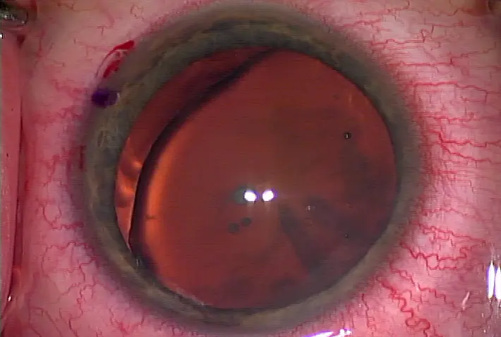
PUK is clearly identifiable in this patient Source: Sophie X. Deng, M.D.

An example of marginal (limbal) herpes simplex keratitis (mimicking PUK) Source: Vincent P. de Luise, M.D.

Mooren's ulcer caused this patient's PUK Source: Vincent P. de Luise, M.D.
Usually considered an ocular manifestation of a systemic autoimmune disorder, peripheral ulcerative keratitis can result in devastating outcomes—including permanent loss of vision
Peripheral ulcerative keratitis (PUK) is typically associated with poorly managed systemic conditions such as rheumatoid arthritis (RA accounts for almost one-third of non-infectious PUK), among others. What can cause it? "VAST CRIMES," said Vincent P. de Luise, M.D., assistant clinical professor of ophthalmology, Yale University School of Medicine, New Haven, Conn. PUK can be caused by "viral, autoimmune, Staphylococcal marginal, Terrien's/furrow degeneration, contact lenses, rosacea, infections beyond viral—such as bacterial, syphilis, Lyme disease, or tuberculosis—Mooren's ulcer, excisional cases—because tumors can mimic—or sclerokeratitis," he said. "It's a useful mnemonic to help run down the list of potential causes." In PUK, the inflammation usually involves the peripheral cornea (as its name suggests), but can also lead to both peripheral and more central corneal melting and perforation if the thinning progresses.
"Almost all the cases I see that are referrals have 3-4 clock hours and are at least 50% thinned," said Jennifer E. Thorne, M.D., chief of the ocular immunology division, and associate professor of ophthalmology and epidemiology, Wilmer Eye Institute, Johns Hopkins University, Baltimore. "They were referred in because the initial attempt to quiet the disease had failed - patients did not respond to corticosteroids and the disease is progressing." Her advice to anyone unlikely to be treating or uncomfortable treating PUK—"refer it to a cornea or uveitis specialist with expertise in PUK," she said. Common signs to look for include peripheral ulceration or infiltrates, said Sophie X. Deng, M.D., assistant professor of ophthalmology, cornea and uveitis division, Jules Stein Eye Institute, David Geffen School of Medicine, University of California, Los Angeles. Plus, most patients will complain of severe photophobia even if the PUK is mild. "If you don't see these kinds of patients very often, it's easy to mistake PUK for an inflammatory disease like bad blepharitis or marginal keratitis," Dr. Deng said. When a 2-3 day course of steroids offers no improvement, think beyond typical keratitis and consider PUK, she said.
One helpful general rule of thumb for people with PUK—"it normally doesn't affect younger people, but mostly people around 50-80 years old," Dr. Deng said. "If the patient comes in, notes contact lens wear, inadvertently slept with the lenses in, and is younger—in his/her 30s—I wouldn't think about PUK as the first diagnosis—I'd treat for infectious keratitis." Dr. de Luise agreed and noted herpes simplex also can mimic symptoms of PUK. "Most clinicians are used to seeing central dendrites," he said. "However, marginal (limbal) HSV keratitis can begin as a marginal ulcer mimicking PUK, and throwing steroids at it will only exacerbate the problem."
As with any disease that has numerous potential causes, a complete patient history is imperative. Dr. de Luise said that not only RA but also Wegener's granulomatosis, polyarteritis nodosa, and Crohn's disease can be associated with PUK. Systemic lupus erythematosus is more rarely associated with PUK. In some cases, PUK may be the first inkling a clinician has that a patient has developed an ocular manifestation of a systemic vasculitis. Mooren's ulcer can manifest as PUK, but Mooren's is a diagnosis of exclusion, he said. PUK associated with RA is more common in women, but PUK seen in Mooren's ulcer is slightly more common in men, Dr. de Luise noted.
Treatment plans
For Dr. de Luise, PUK treatment can be remembered with a second mnemonic: "the Triple A—arrest the corneal disease by medical or surgical means if appropriate, add systemic immunosuppression drugs if an underlying systemic disorder is causing the PUK, and alert the rheumatologist the immunosuppression drugs need to be re-evaluated."
Co-management with a rheumatologist is recommended, "especially when you're unsure how extensive the workup should be or uncomfortable with prescribing and managing immunosuppressive drugs," Dr. Thorne said. If a patient is on medication for RA but develops PUK anyway, it's probable an increase in medication is necessary to get the systemic disease under better control, she said. "[Stephen] Foster et al. published a paper in the journal Ophthalmology almost 30 years ago on mortality in cases of PUK or scleritis in the setting of RA," Dr. de Luise said. "If we're wrong in our diagnosis of PUK, the patient will likely not die of the condition. But if we're right—and the patient has an underlying systemic vasculitis that we diagnosed by identifying the PUK as a marker for systemic disease—that patient's life can be saved and prolonged. Any time a PUK case is behaving suspiciously, get serologies —specifically RF [rheumatoid factor], ANA [anti-nuclear antibody] and ANCA [anti-neutrophil cytoplasmic antibody] titers, C-reactive protein, Lyme titer (if the history suggests), FTA-Abs [fluorescent treponemal antibody absorption], and a chest X-ray. Do a corneal scrape and culture for the possibility of a local infectious cause, such as limbal (marginal) HSV."
Co-management is crucial to align the systemic immunomodulators with the topical drops, Dr. Deng said. "PUK means the systemic vasculitis is there and the disease is not controlled. You need the rheumatologist to change the systemic medications to better manage the disease." On occasion, she's given systemic steroids "for inflammation control," also noting the higher mortality rate associated with poorly managed RA/PUK.
"Clinicians can also admit the patient to the hospital for a pulse dose steroid first and monitoring. The patient can be started on other immune modulating agents," she said. "Oral non-steroidals are not very helpful in managing PUK, and topical non-steroidals can rarely lead to corneal melt," Dr. de Luise said. Cyclophosphamide, methotrexate (oral or subcutaneous), azathioprine, and tumor necrosis factor-alpha antagonists (infliximab, rituximab, etanercept) have all been used to effectively manage PUK in co-existent RA, he added. "Always worry about infection in these eyes," Dr. Deng said. "Culture, culture, culture to ensure there's no secondary infection. Only after you're certain there's no additional infection would I recommend systemic steroid first to control the inflammation."
If surgery is deemed necessary, Dr. Deng recommended tissue adhesives post-keratectomy and conjunctival resection, and amniotic membrane transplantation can be helpful in Mooren's ulcer cases. (She's seen cases of 360-degree melt associated with PUK.)
Future surgery concerns
For patients with systemic autoimmune disorders and who have had PUK, "ideally the inflammation should be controlled for at least 3 months before considering cataract surgery," Dr. Thorne said. In this group of patients, surgeons need to be particularly mindful of the area of corneal thinning, she said. "It may affect the method of cataract removal planned. Additionally, surgeons might have some astigmatism to take into account because of the thinning," she said. Be "very careful about wound construction and possible melting," Dr. de Luise said. "Just the trigger of a diamond knife or femto laser can incite ulcerations." When the systemic disease is quiet both locally and systemically, "one can cautiously proceed with cataract or elective anterior segment surgery, but always be vigilant about wound healing issues." For patients undergoing cataract surgery after successfully managed PUK, Dr. de Luise said "the placement of the cataract wound in these cases is even more crucial than in complicated cases, to avoid problems with wound healing." He recommended using a more scleral incision, one that is self-sealing, and under a conjunctival flap, and to avoid clear/near-clear incisions.
"These eyes are at risk for recurrent inflammation and ulceration," he said. Whether or not to use 10-0 nylon has been debated, as the suture may help with wound healing but is another inflammatory stimulus, he added. In PUK, up to "a third of the stroma can already be gone," Dr. de Luise said, and suggested waiting at least 3 months before recommending that patient for anterior segment surgery. "Weeks is too early for surgery post-PUK; ideally you should wait about 6 months," he said.
Dr. Deng said during an acute phase melting may be so severe, she'd recommend putting off surgery as long as possible. If the eye has excessive thinning, it can quickly become ectatic and develop cataract after PUK resolution because of the inflammation, she said. "For these patients, I use systemic steroids perioperatively, then taper the steroids gradually to get them over the stressful period," she said. "Patients do pretty well that way."
The bottom line for these patients—as long as the systemic disease is well controlled, the ocular manifestations will be controlled accordingly, Dr. Deng said.
Editors' note: The doctors mentioned have no financial interests related to this article.
Contact information
de Luise: 203-263-3300, vdeluisemd@gmail.com
Deng: 310-206-7202, deng@jsei.ucla.edu
Thorne: 410-955-2966, jthorne@jhmi.edu
PUK and systemic autoimmune disease
by Michelle Dalton EyeWorld Contributing Editor
PUK is clearly identifiable in this patient Source: Sophie X. Deng, M.D.
An example of marginal (limbal) herpes simplex keratitis (mimicking PUK) Source: Vincent P. de Luise, M.D.
Mooren's ulcer caused this patient's PUK Source: Vincent P. de Luise, M.D.
Usually considered an ocular manifestation of a systemic autoimmune disorder, peripheral ulcerative keratitis can result in devastating outcomes—including permanent loss of vision
Peripheral ulcerative keratitis (PUK) is typically associated with poorly managed systemic conditions such as rheumatoid arthritis (RA accounts for almost one-third of non-infectious PUK), among others. What can cause it? "VAST CRIMES," said Vincent P. de Luise, M.D., assistant clinical professor of ophthalmology, Yale University School of Medicine, New Haven, Conn. PUK can be caused by "viral, autoimmune, Staphylococcal marginal, Terrien's/furrow degeneration, contact lenses, rosacea, infections beyond viral—such as bacterial, syphilis, Lyme disease, or tuberculosis—Mooren's ulcer, excisional cases—because tumors can mimic—or sclerokeratitis," he said. "It's a useful mnemonic to help run down the list of potential causes." In PUK, the inflammation usually involves the peripheral cornea (as its name suggests), but can also lead to both peripheral and more central corneal melting and perforation if the thinning progresses.
"Almost all the cases I see that are referrals have 3-4 clock hours and are at least 50% thinned," said Jennifer E. Thorne, M.D., chief of the ocular immunology division, and associate professor of ophthalmology and epidemiology, Wilmer Eye Institute, Johns Hopkins University, Baltimore. "They were referred in because the initial attempt to quiet the disease had failed - patients did not respond to corticosteroids and the disease is progressing." Her advice to anyone unlikely to be treating or uncomfortable treating PUK—"refer it to a cornea or uveitis specialist with expertise in PUK," she said. Common signs to look for include peripheral ulceration or infiltrates, said Sophie X. Deng, M.D., assistant professor of ophthalmology, cornea and uveitis division, Jules Stein Eye Institute, David Geffen School of Medicine, University of California, Los Angeles. Plus, most patients will complain of severe photophobia even if the PUK is mild. "If you don't see these kinds of patients very often, it's easy to mistake PUK for an inflammatory disease like bad blepharitis or marginal keratitis," Dr. Deng said. When a 2-3 day course of steroids offers no improvement, think beyond typical keratitis and consider PUK, she said.
One helpful general rule of thumb for people with PUK—"it normally doesn't affect younger people, but mostly people around 50-80 years old," Dr. Deng said. "If the patient comes in, notes contact lens wear, inadvertently slept with the lenses in, and is younger—in his/her 30s—I wouldn't think about PUK as the first diagnosis—I'd treat for infectious keratitis." Dr. de Luise agreed and noted herpes simplex also can mimic symptoms of PUK. "Most clinicians are used to seeing central dendrites," he said. "However, marginal (limbal) HSV keratitis can begin as a marginal ulcer mimicking PUK, and throwing steroids at it will only exacerbate the problem."
As with any disease that has numerous potential causes, a complete patient history is imperative. Dr. de Luise said that not only RA but also Wegener's granulomatosis, polyarteritis nodosa, and Crohn's disease can be associated with PUK. Systemic lupus erythematosus is more rarely associated with PUK. In some cases, PUK may be the first inkling a clinician has that a patient has developed an ocular manifestation of a systemic vasculitis. Mooren's ulcer can manifest as PUK, but Mooren's is a diagnosis of exclusion, he said. PUK associated with RA is more common in women, but PUK seen in Mooren's ulcer is slightly more common in men, Dr. de Luise noted.
Treatment plans
For Dr. de Luise, PUK treatment can be remembered with a second mnemonic: "the Triple A—arrest the corneal disease by medical or surgical means if appropriate, add systemic immunosuppression drugs if an underlying systemic disorder is causing the PUK, and alert the rheumatologist the immunosuppression drugs need to be re-evaluated."
Co-management with a rheumatologist is recommended, "especially when you're unsure how extensive the workup should be or uncomfortable with prescribing and managing immunosuppressive drugs," Dr. Thorne said. If a patient is on medication for RA but develops PUK anyway, it's probable an increase in medication is necessary to get the systemic disease under better control, she said. "[Stephen] Foster et al. published a paper in the journal Ophthalmology almost 30 years ago on mortality in cases of PUK or scleritis in the setting of RA," Dr. de Luise said. "If we're wrong in our diagnosis of PUK, the patient will likely not die of the condition. But if we're right—and the patient has an underlying systemic vasculitis that we diagnosed by identifying the PUK as a marker for systemic disease—that patient's life can be saved and prolonged. Any time a PUK case is behaving suspiciously, get serologies —specifically RF [rheumatoid factor], ANA [anti-nuclear antibody] and ANCA [anti-neutrophil cytoplasmic antibody] titers, C-reactive protein, Lyme titer (if the history suggests), FTA-Abs [fluorescent treponemal antibody absorption], and a chest X-ray. Do a corneal scrape and culture for the possibility of a local infectious cause, such as limbal (marginal) HSV."
Co-management is crucial to align the systemic immunomodulators with the topical drops, Dr. Deng said. "PUK means the systemic vasculitis is there and the disease is not controlled. You need the rheumatologist to change the systemic medications to better manage the disease." On occasion, she's given systemic steroids "for inflammation control," also noting the higher mortality rate associated with poorly managed RA/PUK.
"Clinicians can also admit the patient to the hospital for a pulse dose steroid first and monitoring. The patient can be started on other immune modulating agents," she said. "Oral non-steroidals are not very helpful in managing PUK, and topical non-steroidals can rarely lead to corneal melt," Dr. de Luise said. Cyclophosphamide, methotrexate (oral or subcutaneous), azathioprine, and tumor necrosis factor-alpha antagonists (infliximab, rituximab, etanercept) have all been used to effectively manage PUK in co-existent RA, he added. "Always worry about infection in these eyes," Dr. Deng said. "Culture, culture, culture to ensure there's no secondary infection. Only after you're certain there's no additional infection would I recommend systemic steroid first to control the inflammation."
If surgery is deemed necessary, Dr. Deng recommended tissue adhesives post-keratectomy and conjunctival resection, and amniotic membrane transplantation can be helpful in Mooren's ulcer cases. (She's seen cases of 360-degree melt associated with PUK.)
Future surgery concerns
For patients with systemic autoimmune disorders and who have had PUK, "ideally the inflammation should be controlled for at least 3 months before considering cataract surgery," Dr. Thorne said. In this group of patients, surgeons need to be particularly mindful of the area of corneal thinning, she said. "It may affect the method of cataract removal planned. Additionally, surgeons might have some astigmatism to take into account because of the thinning," she said. Be "very careful about wound construction and possible melting," Dr. de Luise said. "Just the trigger of a diamond knife or femto laser can incite ulcerations." When the systemic disease is quiet both locally and systemically, "one can cautiously proceed with cataract or elective anterior segment surgery, but always be vigilant about wound healing issues." For patients undergoing cataract surgery after successfully managed PUK, Dr. de Luise said "the placement of the cataract wound in these cases is even more crucial than in complicated cases, to avoid problems with wound healing." He recommended using a more scleral incision, one that is self-sealing, and under a conjunctival flap, and to avoid clear/near-clear incisions.
"These eyes are at risk for recurrent inflammation and ulceration," he said. Whether or not to use 10-0 nylon has been debated, as the suture may help with wound healing but is another inflammatory stimulus, he added. In PUK, up to "a third of the stroma can already be gone," Dr. de Luise said, and suggested waiting at least 3 months before recommending that patient for anterior segment surgery. "Weeks is too early for surgery post-PUK; ideally you should wait about 6 months," he said.
Dr. Deng said during an acute phase melting may be so severe, she'd recommend putting off surgery as long as possible. If the eye has excessive thinning, it can quickly become ectatic and develop cataract after PUK resolution because of the inflammation, she said. "For these patients, I use systemic steroids perioperatively, then taper the steroids gradually to get them over the stressful period," she said. "Patients do pretty well that way."
The bottom line for these patients—as long as the systemic disease is well controlled, the ocular manifestations will be controlled accordingly, Dr. Deng said.
Editors' note: The doctors mentioned have no financial interests related to this article.
Contact information
de Luise: 203-263-3300, vdeluisemd@gmail.com
Deng: 310-206-7202, deng@jsei.ucla.edu
Thorne: 410-955-2966, jthorne@jhmi.edu


留言
張貼留言