Review of "Effects of tamsulosin and silodosin on isolated albino and pigmented rabbit iris dilators: Possible mechanism of intraoperative floppy-iris syndrome"
Review of "Effects of tamsulosin and silodosin on isolated albino and pigmented rabbit iris dilators: Possible mechanism of intraoperative floppy-iris syndrome"
--------------------------------------------------------------------------------

Steven J. Gedde, M.D.
This month, an important paper on the mechanism of IFIS appears in JCRS. I invited the Bascom Palmer residents to review it, and their analysis will help our clinician readers to better understand the findings and their significance.
—David F. Chang, M.D., EyeWorld chief medical editor

The residents from Bascom Palmer Eye Institute Source: Steven J. Gedde, M.D.
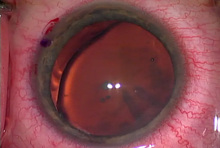
Intraoperative floppy iris syndrome (IFIS) is a recently described condition that may present significant challenges during cataract surgery. IFIS is defined by three intraoperative features: billowing of the iris stroma in response to intraocular microfluidic currents, an increased tendency for the iris to prolapse through the phaco and/or sideport incisions, and an abnormal predisposition toward progressive intraoperative miosis despite standard techniques for pre-op dilation.1,2 IFIS has been strongly associated with use of α1 adrenergic antagonists, medications commonly used to treat benign prostatic hyperplasia. This association is particularly strong with tamsulosin, a long-acting α1A receptor antagonist. The pathophysiology of tamsulosin- mediated IFIS remains poorly understood.
Because of the long half-life of tamsulosin, one hypothesis is that prolonged α1A receptor blockade may lead to chronic inhibition of the iris dilator muscle with resultant "disuse atrophy." Both cadaveric and anterior segment ocular coherence tomography (OCT) studies have demonstrated significantly decreased iris dilator muscle thickness in eyes of patients who previously took tamsulosin compared with age-matched controls, suggesting that chronic tamsulosin use may lead to atrophic anatomical changes in the iris.3,4 Prior studies in patients with IFIS by the authors of the current paper have demonstrated histological changes in iris pigment granules and the iris pigmented epithelium.5 As such, the authors explored the hypothesis that the interactions between α1 antagonists and iris melanin granules may play a crucial role in mediating IFIS.
To study this question, the authors used explanted rabbit iris dilator muscles and measured iris tension in response to phenylephrine without and with an α1 agonist. They first looked at the concentration response curve to phenylephrine in Dutch pigmented rabbits and albino Japanese rabbits. Interestingly, they demonstrated that the concentration response curve for phenylephrine was less right-shifted with higher concentrations of tamsulosin in pigmented rabbit irides as compared to albino irides, and that pigmented rabbit irides had a higher pKb, a measure of affinity for a given receptor, compared to albinos. This illustrates that phenylephrine has a higher binding affinity to the α1 receptor for pigmented as compared to albino irides, suggesting that the presence of melanin pigment may allow increased binding of the α1 receptor. One major assumption not verified in these experiments is that the pigmented and albino rabbit irides are identical except for their melanin content. It is well known that different strains of animals can differ significantly in their physiological function and how they react to drugs from a pharmacokinetic and pharmacodynamic perspective. Additional control experiments examining the distribution and quantity of α1A receptors between pigmented and albino irides would have supported the authors' assumption.
In a second set of experiments, the authors looked at the effect of α1A blockade on iris contraction in an in vivo model by administering daily intramuscular injections of tamsulosin or silodosin over 4 weeks. Of note, there were no albino rabbits utilized in these experiments. The authors noted a statistically significant reduction in maximum dilator contraction after repeated phenylephrine exposure (third and fourth exposure) in dilators receiving α1A antagonists compared to controls. This suggests that there may be some increased fatigability of the iris dilator muscle in tamsulosin-treated animals versus controls. While this decrement in dilator contraction may be attributable to disuse atrophy from α1A blockade as the authors propose, it does not delineate the particular role of the iris pigment granules, as no comparison was done between pigmented and albino rabbits. A more direct experiment to evaluate their hypothesis would be to study differences in response to phenylephrine between pigmented and albino rabbits that have received chronic α1A receptor blockade. The authors also conducted structural analyses using electron microscopy (EM) on rabbit irides that received chronic tamsulosin versus controls. The authors noted irregularities in the size and clumping of pigment granules, thinning of the dilator muscle, and a more lobular, irregular appearance of the shared nuclei of the dilator muscle and pigment epithelium, similar to what had been observed in the human specimens. However, no quantitative data comparing irregularities of pigment clumping and thickness of the dilator muscle was presented to interpret the variability of these morphological features. The authors postulate that α1A antagonist affinity for melanin found in the epithelial cells contributes to dilator muscle atrophy. To provide stronger evidence for their hypothesis, one could perform EM analyses of pigmented and albino rabbit eyes that have been exposed to α1A antagonists. If iris dilator muscles are less susceptible to α1A antagonist-induced atrophy in albino as compared to pigmented rabbits, this would support the authors' hypothesis. Another potential experiment would be to study the dose response relationship of iris dilator muscle atrophy based on increasing exposure to α1 antagonists in heavily versus lightly pigmented irides, using anterior segment OCT to quantitate levels of atrophy over time. Overall, this study makes an important attempt to use experimental animal models to investigate the mechanism behind IFIS. Caution must be used in extrapolating the results of animal studies to human patients. While this study does demonstrate some pharmacologic responses between pigmented and albino irides, additional experiments would help to further define the role of drug pigment interactions in the pathophysiology of IFIS. As mentioned above, direct comparison between pigmented and albino irides in the chronic tamsulosin model with pharmacologic as well as anatomical analyses would be very instructive.
Finally, it is noteworthy that no association between iris color and IFIS was found in a prior prospective clinical study,1 nor is there a body of empiric evidence suggesting that brown-eyed patients are more susceptible to α1 antagonist-induced IFIS than blue-eyed patients. A prospective study of individuals correlating iris color, administration of tamsulosin, and severity of IFIS may help to demonstrate the contribution of drug-melanin interactions in IFIS. Although difficult due to its rarity, patients or animals with unilateral congenital iris heterochromia might be the ideal subjects to test the hypothesis that darker pigmented irides are more susceptible to IFIS by means of α1 antagonist-pigment interaction. We congratulate the authors for this important study that provides information to help us better understand the pathophysiology of IFIS.
References
1. Chang DF, Braga-Mele R, Mamalis N, Masket S, Miller KM, Nichamin LD, Packard RB, Packer M; ASCRS Cataract Clinical Committee. ASCRS White Paper: clinical review of intraoperative floppy-iris syndrome. J Cataract Refract Surg. 2008;34(12):2153-62.
2. Chang DF, Campbell JR. Intraoperative floppy iris syndrome associated with tamsulosin. J Cataract Refract Surg. 2005;31(4):664-73.
3. Santaella RM, Destafeno JJ, Stinnett SS, Proia AD, Chang DF, Kim T. The effect of alpha1-adrenergic receptor antagonist tamsulosin (Flomax) on iris dilator smooth muscle anatomy. Ophthalmology. 2010;117(9):1743-9.
4. Prata TS, Palmiero PM, Angelilli A, Sbeity Z, De Moraes CG, Liebmann JM, Ritch R. Iris morphologic changes related to alpha(1)-adrenergic receptor antagonists implications for intraoperative floppy iris syndrome. Ophthalmology. 2009;116(5):877-81.
5. Goseki T, Shimizu K, Ishikawa H, Nishimoto H, Uga S, Nemoto N, Patil PN. Possible mechanism of intraoperative floppy iris syndrome: a clinicopathological study. Br J Ophthalmology. 2008; 92(8):1156-8.
Contact information
Gedde: sgedde@med.miami.edu
--------------------------------------------------------------------------------
by Daniel L. Chao, M.D., Yasha S. Modi, M.D., Bradford Lee, M.D., Jayanth Sridhar, M.D., Ajay E. Kuriyan M.D., and Steven J. Gedde, M.D., Bascom Palmer Eye Institute, Miami, Fla.
Steven J. Gedde, M.D.
This month, an important paper on the mechanism of IFIS appears in JCRS. I invited the Bascom Palmer residents to review it, and their analysis will help our clinician readers to better understand the findings and their significance.
—David F. Chang, M.D., EyeWorld chief medical editor
The residents from Bascom Palmer Eye Institute Source: Steven J. Gedde, M.D.
Intraoperative floppy iris syndrome (IFIS) is a recently described condition that may present significant challenges during cataract surgery. IFIS is defined by three intraoperative features: billowing of the iris stroma in response to intraocular microfluidic currents, an increased tendency for the iris to prolapse through the phaco and/or sideport incisions, and an abnormal predisposition toward progressive intraoperative miosis despite standard techniques for pre-op dilation.1,2 IFIS has been strongly associated with use of α1 adrenergic antagonists, medications commonly used to treat benign prostatic hyperplasia. This association is particularly strong with tamsulosin, a long-acting α1A receptor antagonist. The pathophysiology of tamsulosin- mediated IFIS remains poorly understood.
Because of the long half-life of tamsulosin, one hypothesis is that prolonged α1A receptor blockade may lead to chronic inhibition of the iris dilator muscle with resultant "disuse atrophy." Both cadaveric and anterior segment ocular coherence tomography (OCT) studies have demonstrated significantly decreased iris dilator muscle thickness in eyes of patients who previously took tamsulosin compared with age-matched controls, suggesting that chronic tamsulosin use may lead to atrophic anatomical changes in the iris.3,4 Prior studies in patients with IFIS by the authors of the current paper have demonstrated histological changes in iris pigment granules and the iris pigmented epithelium.5 As such, the authors explored the hypothesis that the interactions between α1 antagonists and iris melanin granules may play a crucial role in mediating IFIS.
To study this question, the authors used explanted rabbit iris dilator muscles and measured iris tension in response to phenylephrine without and with an α1 agonist. They first looked at the concentration response curve to phenylephrine in Dutch pigmented rabbits and albino Japanese rabbits. Interestingly, they demonstrated that the concentration response curve for phenylephrine was less right-shifted with higher concentrations of tamsulosin in pigmented rabbit irides as compared to albino irides, and that pigmented rabbit irides had a higher pKb, a measure of affinity for a given receptor, compared to albinos. This illustrates that phenylephrine has a higher binding affinity to the α1 receptor for pigmented as compared to albino irides, suggesting that the presence of melanin pigment may allow increased binding of the α1 receptor. One major assumption not verified in these experiments is that the pigmented and albino rabbit irides are identical except for their melanin content. It is well known that different strains of animals can differ significantly in their physiological function and how they react to drugs from a pharmacokinetic and pharmacodynamic perspective. Additional control experiments examining the distribution and quantity of α1A receptors between pigmented and albino irides would have supported the authors' assumption.
In a second set of experiments, the authors looked at the effect of α1A blockade on iris contraction in an in vivo model by administering daily intramuscular injections of tamsulosin or silodosin over 4 weeks. Of note, there were no albino rabbits utilized in these experiments. The authors noted a statistically significant reduction in maximum dilator contraction after repeated phenylephrine exposure (third and fourth exposure) in dilators receiving α1A antagonists compared to controls. This suggests that there may be some increased fatigability of the iris dilator muscle in tamsulosin-treated animals versus controls. While this decrement in dilator contraction may be attributable to disuse atrophy from α1A blockade as the authors propose, it does not delineate the particular role of the iris pigment granules, as no comparison was done between pigmented and albino rabbits. A more direct experiment to evaluate their hypothesis would be to study differences in response to phenylephrine between pigmented and albino rabbits that have received chronic α1A receptor blockade. The authors also conducted structural analyses using electron microscopy (EM) on rabbit irides that received chronic tamsulosin versus controls. The authors noted irregularities in the size and clumping of pigment granules, thinning of the dilator muscle, and a more lobular, irregular appearance of the shared nuclei of the dilator muscle and pigment epithelium, similar to what had been observed in the human specimens. However, no quantitative data comparing irregularities of pigment clumping and thickness of the dilator muscle was presented to interpret the variability of these morphological features. The authors postulate that α1A antagonist affinity for melanin found in the epithelial cells contributes to dilator muscle atrophy. To provide stronger evidence for their hypothesis, one could perform EM analyses of pigmented and albino rabbit eyes that have been exposed to α1A antagonists. If iris dilator muscles are less susceptible to α1A antagonist-induced atrophy in albino as compared to pigmented rabbits, this would support the authors' hypothesis. Another potential experiment would be to study the dose response relationship of iris dilator muscle atrophy based on increasing exposure to α1 antagonists in heavily versus lightly pigmented irides, using anterior segment OCT to quantitate levels of atrophy over time. Overall, this study makes an important attempt to use experimental animal models to investigate the mechanism behind IFIS. Caution must be used in extrapolating the results of animal studies to human patients. While this study does demonstrate some pharmacologic responses between pigmented and albino irides, additional experiments would help to further define the role of drug pigment interactions in the pathophysiology of IFIS. As mentioned above, direct comparison between pigmented and albino irides in the chronic tamsulosin model with pharmacologic as well as anatomical analyses would be very instructive.
Finally, it is noteworthy that no association between iris color and IFIS was found in a prior prospective clinical study,1 nor is there a body of empiric evidence suggesting that brown-eyed patients are more susceptible to α1 antagonist-induced IFIS than blue-eyed patients. A prospective study of individuals correlating iris color, administration of tamsulosin, and severity of IFIS may help to demonstrate the contribution of drug-melanin interactions in IFIS. Although difficult due to its rarity, patients or animals with unilateral congenital iris heterochromia might be the ideal subjects to test the hypothesis that darker pigmented irides are more susceptible to IFIS by means of α1 antagonist-pigment interaction. We congratulate the authors for this important study that provides information to help us better understand the pathophysiology of IFIS.
References
1. Chang DF, Braga-Mele R, Mamalis N, Masket S, Miller KM, Nichamin LD, Packard RB, Packer M; ASCRS Cataract Clinical Committee. ASCRS White Paper: clinical review of intraoperative floppy-iris syndrome. J Cataract Refract Surg. 2008;34(12):2153-62.
2. Chang DF, Campbell JR. Intraoperative floppy iris syndrome associated with tamsulosin. J Cataract Refract Surg. 2005;31(4):664-73.
3. Santaella RM, Destafeno JJ, Stinnett SS, Proia AD, Chang DF, Kim T. The effect of alpha1-adrenergic receptor antagonist tamsulosin (Flomax) on iris dilator smooth muscle anatomy. Ophthalmology. 2010;117(9):1743-9.
4. Prata TS, Palmiero PM, Angelilli A, Sbeity Z, De Moraes CG, Liebmann JM, Ritch R. Iris morphologic changes related to alpha(1)-adrenergic receptor antagonists implications for intraoperative floppy iris syndrome. Ophthalmology. 2009;116(5):877-81.
5. Goseki T, Shimizu K, Ishikawa H, Nishimoto H, Uga S, Nemoto N, Patil PN. Possible mechanism of intraoperative floppy iris syndrome: a clinicopathological study. Br J Ophthalmology. 2008; 92(8):1156-8.
Contact information
Gedde: sgedde@med.miami.edu


留言
張貼留言